MR Enterography Services

ABOUT MR Enterography at MetroWest MRI

MR Enterography is a magnetic resonance imaging (MRI) technique that doctors use to evaluate the small intestine. MRI uses a safe magnetic field and radio waves to create images of the body. MR Enterography does not expose you to any radiation.

Doctors use MR Enterography to diagnose and evaluate conditions such as:

  • Inflammation
  • Crohn’s disease and ulcerative colitis
  • Infection
  • Tumors
  • Abscesses and fistulas
  • Bowel obstructions

Patient information

Prior to your exam, review the following checklist:

  • Do not eat or drink for six hours prior to your MR Enterography.
  • Continue to take any medication you normally take.
  • If you may be claustrophobic, ask your doctor to prescribe a mild sedative prior to the exam. If you do receive medication, bring someone with you to drive you home.

At the clinic, you will drink three bottles of Volumen, a water-based solution to distend the small bowel. You will do this over the course of 60 minutes.

The MRI scan will then take 30 – 60 minutes. During the exam, you will receive an injected contrast agent. MRI contrast agents are safe for most people. If you have a history of kidney disease, your doctor may test your kidney function prior to the exam.

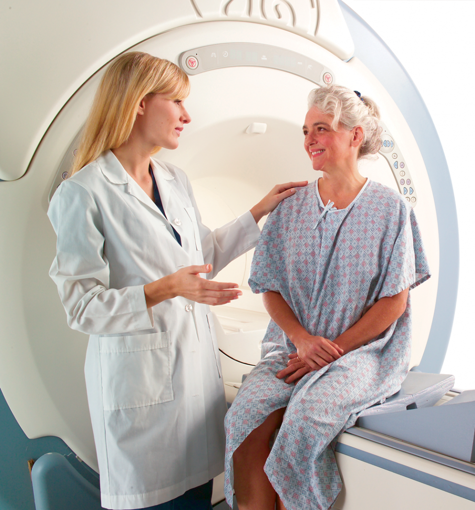
The technologist stays in contact with you throughout the exam.

After your exam is complete, a board-certified radiologist will review the scans and forward results to your doctor.